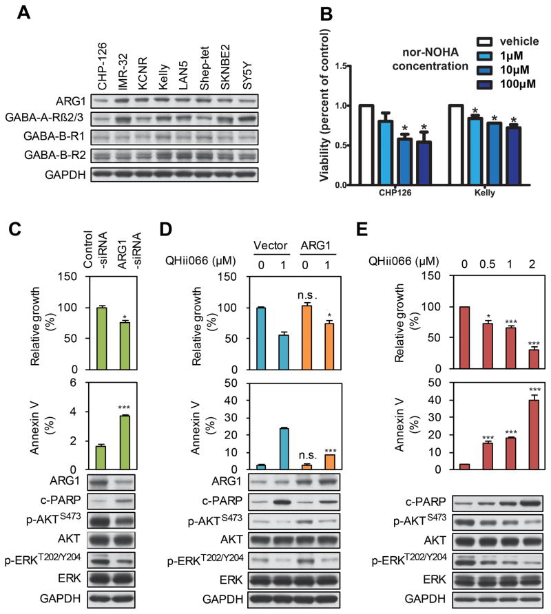
Figure 5. ARG1 inhibition and GABA activation suppress neuroblastoma cell growth and survival.
(A) Expression of ARG1, GABA-A, and GABA-B receptors in neuroblastoma cell lines. Cell lines as indicated were harvested, lysed and analyzed by Western blot using the antisera indicated. (B) WST-1 assay showing a dose-dependent decrease in viability following 72h treatment of neuroblastoma cells with varying doses of the ARG1 inhibitor nor-NOHA. (C) LAN5 cells were transfected with either control or ARG1 siRNA. Cell viability was measured by WST-1. Apoptosis was measured by flow cytometry for the apoptotic marker annexin V. An aliquot of cells was analyzed by immunoblot using antisera indicated. (D) LAN-5-pBABE-vector and LAN-5-pBABE-ARG1 cells were treated with DMSO or with the GABA-A agonist QHii066. Cell viability, apoptosis, and protein markers were measured as in panel (C). (E) LAN-5 cells were treated with the GABA-A agonist QHii066 for 3 days. Cell viability, apoptosis, and protein markers were measured as in panel (C). For panels B–E, data shown are mean ± SD of triplicate measurements. NS, not significant vs control/vehicle using student’s t-test; * indicates p<0.05; *** indicates p<0.001.

