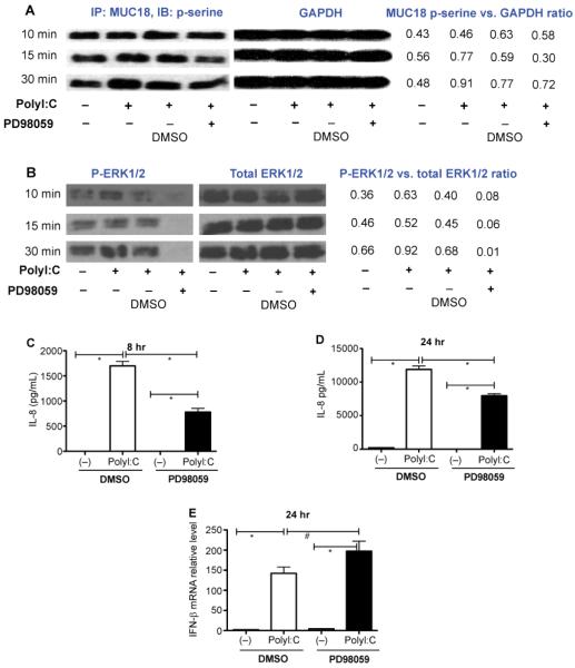
Figure 4.
MUC18 serine phosphorylation in polyI:C-stimulated human lung epithelial cell line NCI-H292. (A) At the indicated time points, cells were immunoprecipitated with a rabbit anti-human MUC18 antibody, and then immunoblotted with an antibody against phosphorylated serines. GAPDH immunoblot was performed using 30 μg total proteins under different conditions to verify equal loading. Densitometry was performed to measure the intensity of MUC18 p-serine and GAPDH signals. The ratio of MUC18 P-serine versus GAPDH was used to indicate the level of phosphorylated MUC18. (B) Cell lysates were immunoblotted with antibodies against phosphorylated ERK1/2 (p-ERK) and total ERK1/2. The ratio of p-ERK1/2 versus total ERK1/2 intensity as evaluated by densitometry was used to indicate the level of phosphorylated ERK1/2. (C–D) ERK inhibition by PD98059 significantly reduced IL-8 production in NCI-H292 cells at 8 hours and 24 hours after polyI:C stimulation. However, PD98059 did not inhibit IFN-β mRNA expression at 24 hours (E). N=12 replicates. *, p<0.05; #, p>0.05.

