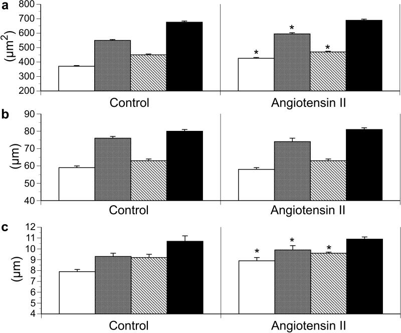
Figure 1.
Effect of angiotensin II infusion on fetal ventricular cardiomyocyte (a) cell area, (b) cell length, and (c) cell width in left ventricle mononucleated cells (white bars); left ventricle binucleated cells (grey bars); right ventricle mononucleated cells (diagonal stripe bars); and right ventricle binucleated cells (black bars). Values expressed as means ± SE. *p <0.05 compared to control of similar cell type.

