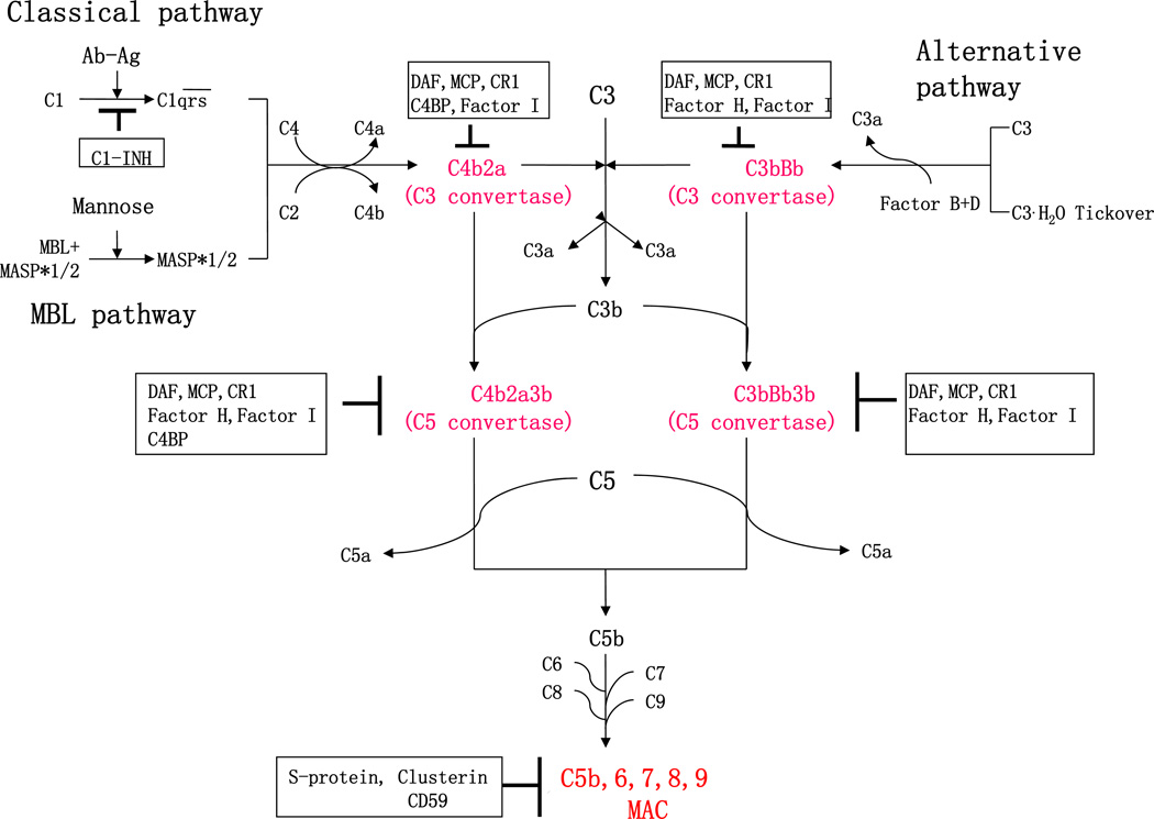
Figure 1. Complement activation and regulation.
The complement system is activated through three different activation pathways: classical, alternative and MBL cascades. The three complement activation pathways converge at the C3 level. Formation of C3 or C5 convertases as well as the membrane attack complement (MAC), a final activation complex (as highlighted in red text) are regulated by a number of complement regulators (highlighted within black squares). Ab, Antibody; Ag, Antigen; C1-INH, C1-inhibitor; MBL, Mannose-binding lectin; MASP, MBL-associated serine proteases; DAF, decay-accelerating factor; MCP, membrane co-factor protein; CR, complement receptor; C4BP, C4b-binding protein; and MAC, membrane attack complex.

