
Figure 8.
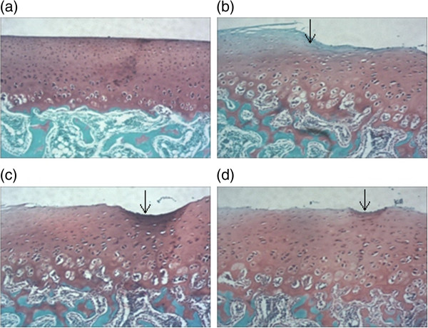
Photographs of rabbit articular cartilage stained by Safranin-O. (a). Normal articular cartilage showed no reduction of Safranin staining. (b). Vehicle-treated cartilage showed the loss of Safranin O staining, the arrow indicates the calcified cartilage and irregular surface. (c),(d) biochanin A -treated cartilage showed reduction loss of Safranin O staining compared with vehicle-treated group (original magnification × 40), the arrow indicate irregular surface. a, b, c, d in Figure 8 represented as normal cartilage, vehicle-treated cartilage, biochanin A (5 μM) and biochanin A (50 μM)-treated cartilage respectively.