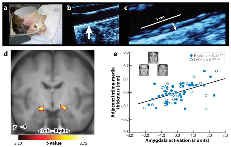
Figure 3.
In a functional neuroimaging study, a measure of preclinical atherosclerosis was related to activation of the amygdala in response to threatening facial expressions. Carotid intima-media thickness (IMT) was assessed by B-mode ultrasonography. (a) A trained vascular technologist imaged the carotid arteries. (b) An image of the carotid artery with the common carotid segment visible at right and the beginning of the carotid bulb visible at left; arrow indicates a point along the far wall of the common carotid artery, which is shown at higher magnification in panel c. (c) Magnified point of the far wall of the common carotid artery illustrating the lumen-intima and media-adventitia interfaces. IMT was measured by averaging the IMT (the distance between two lines tracking the lumen-intima and media-adventitia interfaces, not illustrated here) in 1-mm increments along the distal 1 cm of the far wall of the common carotid artery, the far wall of the carotid bulb, and the first centimeter of the internal carotid artery. As assessed by this ultrasound procedure, carotid IMT covaried positively with amygdala reactivity to angry and fearful facial expressions in an adult sample of otherwise healthy humans. (d) Statistical parametric maps from a regression analysis identifying regions of the left and right amygdala where carotid IMT covaried with amygdala reactivity after controlling for age, sex, resting systolic blood pressure, and family income. (e) Adjusted IMT values are shown as a function of amygdala activation values of the left (open circles) and right (closed circles) amygdala areas profiled in panel d. Insets in panel e illustrate sample trials from the facial-expression protocol designed to elicit amygdala reactivity. **p < 0.001.

