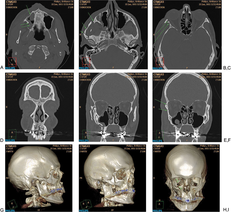
Figure 2.

Systematic analysis of a midfacial fracture. (A) Axial slice: evaluation of the lower central midface with fracture of the zygomatic alveolar crest, anterior and dorsolateral maxillary sinus wall (arrows). (B) Axial slice: Involvement of the zygomatic arch with multiple fractures (arrows). (C) Axial Slice: Fracture of the anterior part of the lateral wall (arrows). (D) Two-dimensional coronal reconstruction at level frontogygomatic buttress (no fracture). (E, F) Two- dimensional coronal reconstruction with the fractures at the zygoma and anterior part of the lateral orbital wall and fronto-zygomatic suture (arrows). (G–I) Three-dimensional reconstruction showing the involvement of the right zygoma, intermediate and lower central midface and orbit.
