Figure 2.
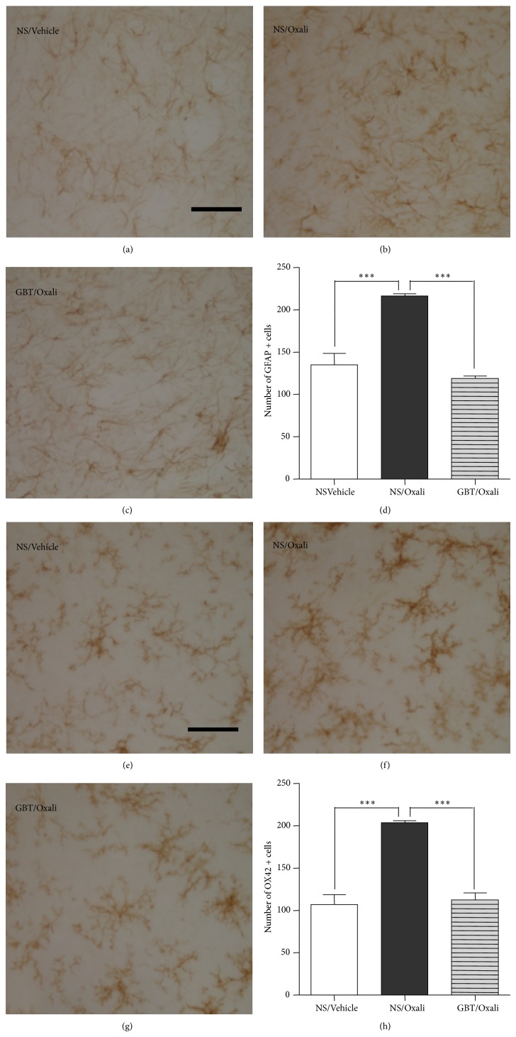
Suppressive effects of GBT on the activation of spinal glial cells. Immunohistochemical analysis of spinal dorsal horn laminae I–IV revealed a morphological activation (b) and an increase in the density (d) of GFAP-positive cells (astrocytes) by an oxaliplatin (6 mg/kg., i.p.) injection, compared to those in a vehicle-injected group (a, d). Such oxaliplatin-induced activation of spinal astrocytes was restored by GBT (400 mg/kg/day) treatments (c, d). Immunohistochemical analysis of spinal dorsal horn also showed a morphological activation (f) and an increase in the density (h) of OX42-positive cells (microglia) by an oxaliplatin injection, compared to those in a vehicle-injected group (e, h). Such microglial activation was restored by GBT treatments (g, h). Data are presented as mean ± S.E.M.*** P < 0.001 by one-way ANOVA followed by Dunnett's post hoc test. Scale bar, 50 μm.

