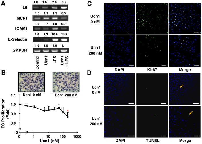
Figure 1. Suppressive effects of Ucn1 on inflammatory response and cell proliferation in human ECs.
(A) HUVECS were pre-treated with or without Ucn1 (200 nmol/l) for 30 min and then incubated with Ucn1 (200 nmol/l)+LPS (1 µg/ml) for 2 h. The mRNA expressions of IL6, MCP1, ICAM1, and E-selectin were analyzed by RT-PCR. GAPDH served as a loading control. Data are representative of 2 independent experiments. (B) Proliferation of EA.hy926 cells was determined by WST-8 assay after 48-h incubation in conditioning medium with the indicated concentrations of Ucn1. Data are expressed as means ± SEM from 4 independent experiments. *P<0.05 vs. 0 nmol/l of Ucn1. (B–D) EA.hy926 cells were stained with Hematoxylin Eosine, anti-Ki-67 antibody, and TUNEL method. DAPI was used to stain the nucleus. Representative images are shown. Scale bar = 100 µm.

