
Figure 2.
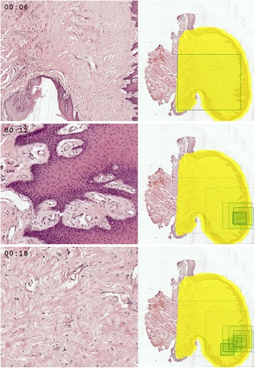
Three frames from an animation showing how a student viewed a WSI with ‘irritation’ fibroma. Left side of each frame presents a fragment displayed on student’s screen at the given moment, while right side shows a WSI thumbnail with positions of the current and previous view fields marked as green rectangles.