
Figure 3.
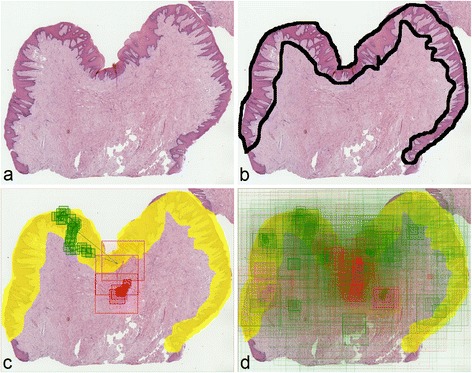
Visualization steps for a question containing a WSI with benign reactive keratosis. (a) Slide overview. (b) Region of interest (diagnostic area) marked by a pathologist for analysis purposes. (c) View paths for two answers (correct = green and incorrect = red), coming from two students. (d) Aggregated map of view paths from all students’ answers for this question.