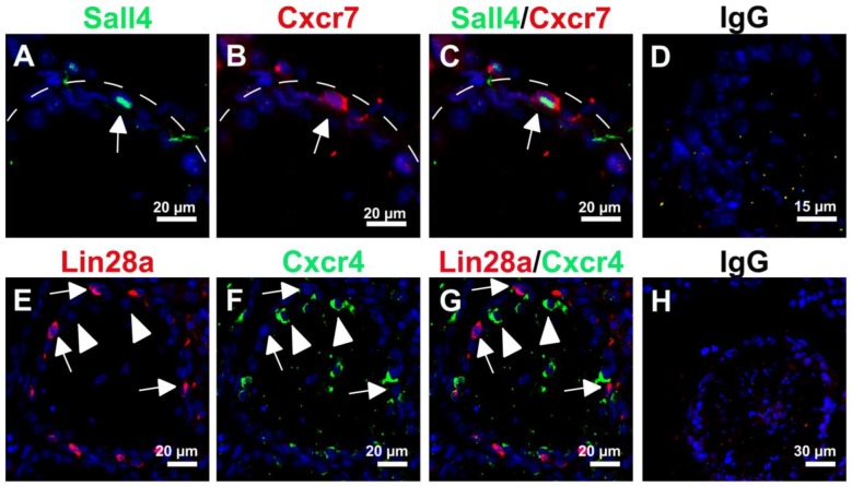
Figure 5. Cxcr7 and Cxcr4 expression in germ cell-depleted mouse testes.
Representative immunofluorescence images of testicular tissue section 28 days after busulfan treatment of adult mice. Cxcr7 expression (B, red, arrow) is restricted to Sall4-positive spermatogonia (A, green, arrow) but Cxcr4 expression (F, green, arrowheads) is not solely observed in Lin28-positive spermatogonia (E, red, arrows). The respective merged images are shown in (C) and (G). Hoechst (blue) was used as nuclear counterstain. Incubation with corresponding IgG antibodies was used as negative control and a representative image is shown in (D, H). Scale bars represent 15, 20 and 30 µm.

