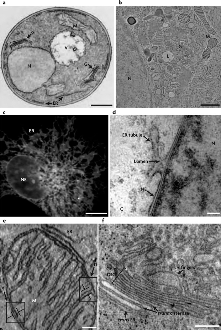
Figure 1.
Simple and complex shapes seen in cells and cell organelles. Image from [3] (Reprinted by permission from Macmillan Publishers Ltd: Nat. Rev. Mol. Cell Biol. 8(3), 258–264 copyright (2007)). (a,b) Electron microscope images showing various cell organelles in a yeast cell (a) and in a normal rat kidney cell (b). The major organelles shown—namely, the Golgi(G), the nucleus(N), the endoplasmic reticulum(ER), the vacuole(V), and the mitochondrion(M)—have identical shapes in both yeast and normal rat kidney cells, and the shapes of these organelles are also preserved in other cells. (c) Fluorescence image of the nuclear envelope(NE) and ER in COS cells, wherein the bright spots denote the localization of a particular ER associated protein. (d) The nuclear envelope that defines the boundary of a cell nucleus is a relatively smooth, double bilayer structure which merges with the rough and tubular endoplasmic reticulum. (e) Doubled walled structure of the mitochondrion; its outer membrane has a smooth shape while its inner membrane is organized into a complex network of tubular shapes called the cristae. (f) Tubules and flattened sacs—the primary structures that dominate the morphology of the Golgi stacks.

