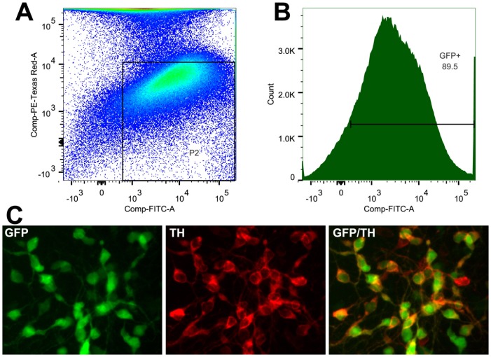
Figure 4. FACS sorting of midbrain DA neurons from E14.5 hTH-GFP rats of Line 12141.
(A) Raw data of sorted cells. Gating for live GFP+ cells was established by utilizing positive controls for GFP and propidium iodide (PI) stained cells (not shown). The gate, P2, excluded weakly GFP+ cells and any PI+ (dead) cells. This allowed for the collection of approximately 90% of GFP+ cells (B). Collected cells were plated at a density of 300,000cells/well and grown for 3 days, fixed and immunostained for TH (C) exhibit similar co-labeling seen in vivo (Figure 1).

