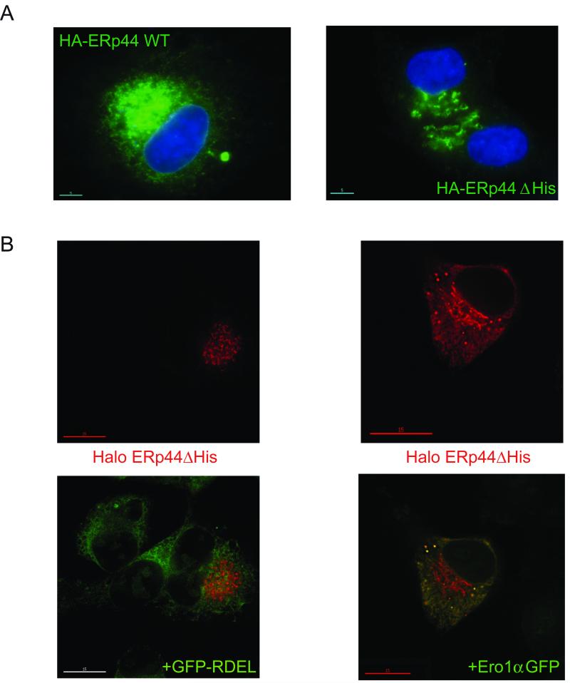
Figure 3. Deletion of histidine rich loop favours accumulation of ERp44 in distal ESC stations.
A) Steady state localization. HepG2 transfectants expressing HA-tagged wt and ΔHis ERp44 were analyzed by immunofluorescence with anti-HA (green). The blue staining (DAPI) was used to detect nuclei. Note that much less green staining is detected in the nuclear membrane or peripheral ER in ΔHis transfectants, as compared to cells expressing wt ERp44.
B) Client-induced relocalisation of ΔHis to the ER. Co-expressing Ero1α-GFP, but not sGFP-RDEL, causes the re-localization of ΔHis in the ER. HepG2 cells were co-transfected with Halo-tagged ERp44 ΔHis and sGFP-RDEL or Ero1α-GFP and then decorated with a Halo ligand (red signal). Clearly, no co-localization is detectable between sGFP-RDEL (green) and ERp44 ΔHis (red) while expression of Ero1α-GFP causes ΔHis to localize also in the ER yielding a reticular yellow staining (right panel).

