Figure 6.
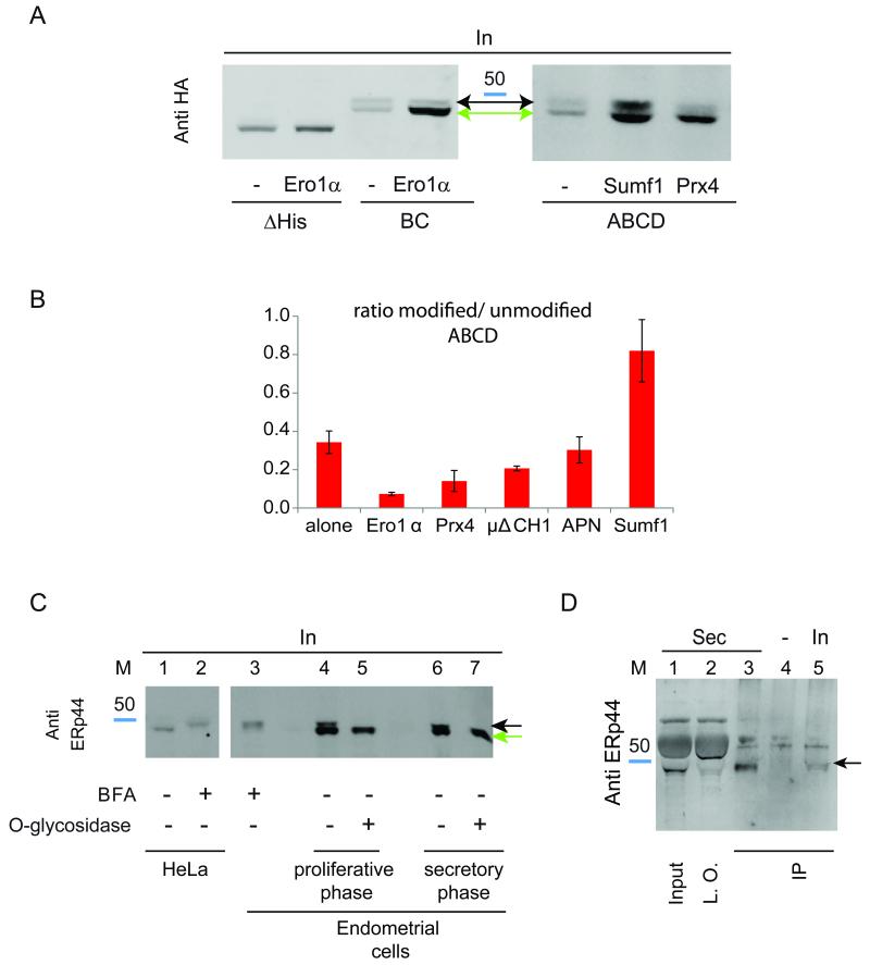
A) Sumf1 can bind ERp44 also after O-glycosylation. HeLa cells were co-transfected as indicated. Aliquots from the lysates were then resolved by SDS-PAGE and blots decorated with anti-HA. Note the accumulation of non-glycosylated histidine mutants in the lysates of cells co-expressing Ero1α or Prx4. Instead, Sumf1 expression allows significant accumulation of O-glycosylated ABCD ERp44.
B) Sequential client-induced retrieval of ERp44 histidine mutants. The histograms show the ratio between modified and unmodified ABCD ERp44 in the presence of Ero1α, Prx4, Sumf1, Ig-μΔCH1 or adiponectin (APN). Data represent the average of ≥3 experiments ± s.d.
C) Endogenous ERp44 is O-glycosylated in primary endometrial cells Aliquots of the lysates obtained from endometrial cells in their proliferative (lanes 4-5) or secretory (lanes 3, 6-7) phase (Di Blasio et al., 1995) were digested with (lanes 5, 7) or without (lanes 3, 4, 6) O-glycosidase and resolved by gel electrophoresis under reducing conditions. Note that upon digestion, the doublet visible in untreated samples collapses into a single higher mobility band, which comprises un-glycosylated and de-glycosylated ERp44 (green arrow). The black arrow points at the O-glycosylated species, the main accumulating in HeLa (lane 2) or endometrial (lane 3) cells after treatment with BFA.
D) Endogenous ERp44 is secreted by endometrial stromal cells Aliquots from the lysates or spent media (48 h) corresponding to 8x10⁴ or 7x10⁵ endometrial cells in their secretory phase were immune-precipitated with 36C9 monoclonal anti ERp44 (IP), resolved under reducing conditions and decorated with rabbit anti ERp44 (JDA1). Lanes 1 and 2 show one third of the spent medium before (Input) or after (left over) immunoprecipitation. The band migrating above the 50 kDa marker is bovine serum albumin, most abundant in foetal calf serum. Cross-linked beads were used as a negative control (lane 4). Endogenous ERp44 is clearly detectable in the culture medium and migrates with a molecular weight similar to the O-glycosylated ERp44 species present intracellularly (black arrow).

