
FIG 2 .
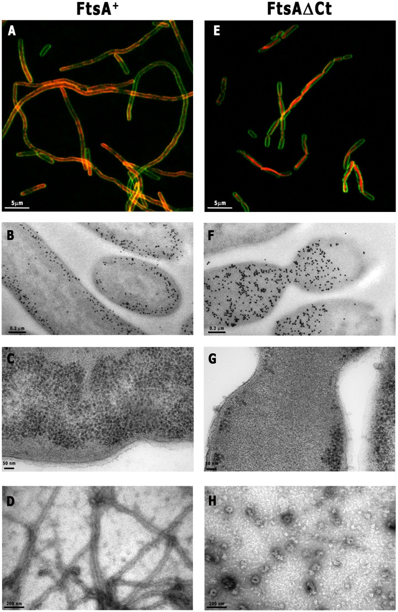
Localization of streptococcal FtsA+ and FtsAΔCt overproduced in E. coli and on lipid monolayers. FtsA (A to C) and FtsAΔCt (E to G) were fused with mCherry (red) at the N terminus and overproduced in E. coli (3 h). Membranes were stained with FM1-43FX (green). Samples were analyzed by confocal microscopy (A and E) and electron microscopy (B, C, F, and G). Panels B and F show the gold immunolocalization of FtsA and FtsAΔCt, respectively. For monolayer assay, a lipid-coated electron microscopy grid was incubated with FtsA+ or FtsAΔCt, followed by the addition of 2 mM ATP (D or H, respectively). Samples were washed extensively with polymerization buffer and fixed with 2% uranyl acetate (see Materials and Methods). (H) Note that polymers formed by FtsAΔCt occurred very occasionally on the lipid monolayer. Scale bars are as indicated.