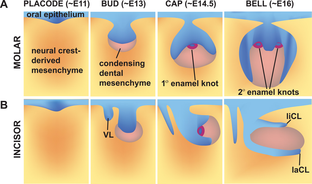
Figure 3. Schematic depiction of mouse molar (a) and incisor (b) tooth development.
Tooth development begins with thickening and invagination of the oral epithelium into the underlying mesenchyme at ~E11. At the bud stage (~E13), the mesenchyme condenses. At the cap stage (~E14.5), the enamel knot, a central signaling center, appears. At the bell stage (~E16), the secondary enamel knots, corresponding to the future location of cusps, form. In addition, the extracellular matrices of enamel and dentin are excreted by the differentiating ameloblasts and odontoblasts, respectively. Tooth development is similar in the incisor and molar, with a few key differences being the proximal-distal “rotation” of the incisor at the bell stage, as well as the absence of secondary enamel knots in the incisor. VL, vestibular lamina.

