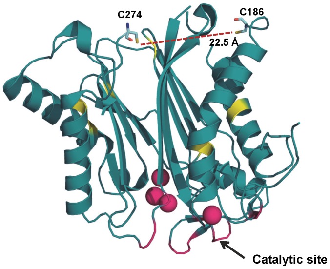
Figure 4. Crystal structure of HAB1.
Ribbon representation of the HAB1 phosphatase domain (PDB: 4LA7) with C186 and C274 presented as stick models and the remaining cysteine residues as yellow patches. The SnRK2.6 interacting site is shown in magenta and MgCl2 ions as magenta spheres (figure drawn from).

