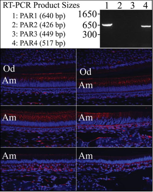
Figure 2.

F2r analyses. Top: RT-PCR of PAR1–PAR4 transcripts from enamel organ epithelia (EOE) of Day 7 mouse molars. PCR conditions were: annealing 58 °C for 30 s; extention at 72 °C for 1 min; 25 cycles. The primers used were: PAR1, F: ctcctcaaggagcagaccac, R: tgcagggactaatgggattc; PAR2, F: tgtgattggtttgcccagta, R: tcgtgacaggtggtgatgtt; PAR3, F: ttctgccagtcactgtttgc, R: ctcgccaaatacccagttgt; PAR4, F: gctggtgctgcactattcaa, R: cacatagcccagcctagctc. Bottom: PAR1 Immunohistochemistry on D14 mandibular incisor. Key: Am, ameloblasts; Od, odontoblasts.
