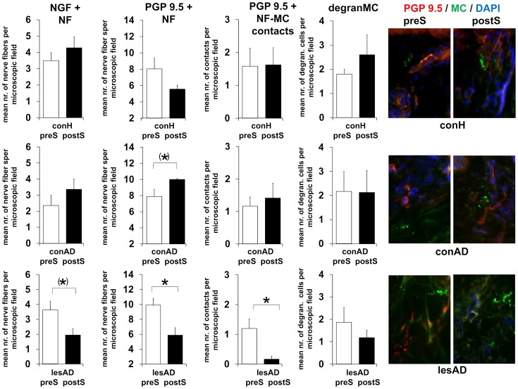
Figure 4. TSST induced neuro-immune changes in healthy control, non-lesional and lesional AD skin.
Changes to baseline obtained as described in Figure 1 were determined 24 h after TSST. Number of participants per evaluation (NGF+ NF, PGP 9.5+ NF, PGP 9.5+ NF-mast cell contacts, degranulated mast cells [MC]): control: N = 11, 4, 4, 4; non-lesional AD: N = 11, 4, 4, 4; lesional AD: N = 11, 6, 6, 5. Corresponding raw data is provided as Data S1. P-values: <0.10 = (*), <0.05 = *. Abbreviations: conH – healthy control skin, conAD – non-lesional AD skin, lesAD – lesional AD skin, preS – prior to TSST, postS – 24 h after TSST.

