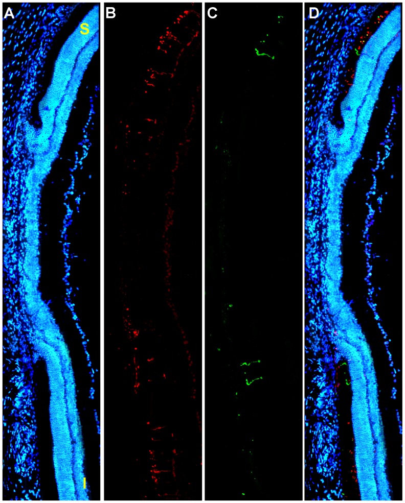
Figure 8. Radial parasagittal section of the retina illustrates focal damage.
A–D. LIP results in focal damage to the outer retina. A. Photomontage of a representative parasagittal oriented radial section spanning the phototoxic lesion stained with DAPI (blue signal) to identify all cell nuclei, from the left retina of an adult albino rat 7 days after LIP. The section was also stained for S- and L-opsins. Note the focal region of damage that shows an absence of the outer nuclear and outer segment layers and results in massive loss of photoreceptors. B,C. L-opsin (red signal) (B) and S-opsin (green signal) (C). Outside the focal damage there are both L- and S-cones, while within the focal damaged region there is only some residual S- or L-immunoreactivity present in the outer segment layer of the retina. D. Couple images with nuclear staining DAPI (blue). S, superior. I, inferior. Bar: 500 µm.

