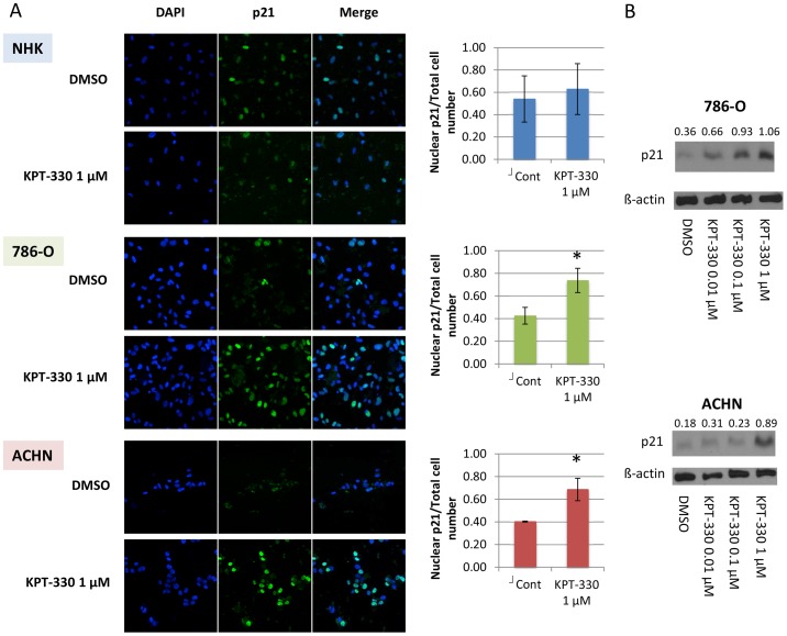
Figure 2. XPO1 inhibition by KPT-330 confined p21 in the nucleus in RCC cells but not in NHK cells.
The RCC cells 786-O and ACHN were grown to ∼60% confluence and treated with KPT-330 at indicated doses for 24 hours. Immunofluorescence was performed by confocal microscopy (20x) and the number of cells in which p21 was predominantly in the nucleus were counted in three randomly selected fields and divided by the total cell number (A). Immunoblotting was accomplished with specific antibodies (B). Blue, nucleus (DAPI); Green, p21. *P<0.05 compared to control. Error bars indicate standard deviation.

