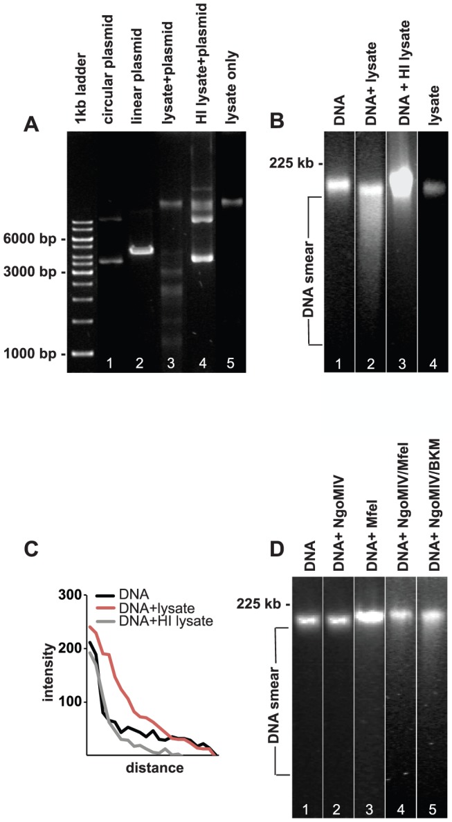
Figure 2. Lysates of N. gonorrhoeae fragments pECFP-N1 and damage DNA from VK2/E6E7 cells.

A. DNA agarose gel showing the digestion of pECFP-N1 plasmid by HindIII (positive control, lane 2), MS11 P+ lysate (lane 3), and MS11 P+ HI lysate (lane 5). Lane 5 shows bacterial MS11 P+ lysate without pECFP-N1 and lane 1 shows uncut circular pECFP-N1. B. PFGE analysis of purified VK2/E6E7 genomic DNA treated for 24 h with: lane 1: PBS (negative control), lane 2: MS11 P+ lysate, lane 3: MS11 P+ HI lysate. Lane 4 shows bacterial MS11 P+ lysate without VK2/E6E7 genomic DNA. C. Graph showing quantification of DNA smears (measured directly underneath and below the band). Shown are smear pixel intensities of cellular DNA alone and cellular DNA exposed to bacterial lysates and HI bacterial lysates. D. PFGE showing genomic DNA subjected to commercial restriction enzymes for 24 h. Lane 1: DNA incubated with CutSmart reaction buffer (negative control). Lane 2: DNA incubated with NgoMIV. Lane 3: DNA incubated with MfeI, Lane 4: DNA incubated with NgoMIV and MfeI Lane 5: DNA incubated with NgoMIV and BamHI/KpnI/MfeI (BKM).
