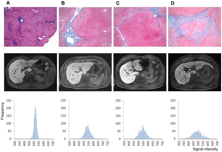
Figure 4. Representative cases of (A) 20 year old male (living-related healthy liver transplantation donor, F0), (B) 37 year old male (hepatocellular carcinoma, F2), (C) 57 year old male (hepatocellular carcinoma, F4), and (D) 54 year old male (hepatocellular carcinoma, F4).
Histology images of Eosin and Masson-Trichrome stain, ×40 (upper row), hepatobiliary phase images of gadoxetic acid-enhanced MRI (middle row), and the histogram produced by plotting each pixel acquired from all four ROI sets according to signal intensity (x-axis) and frequency (y-axis) (lower row).

