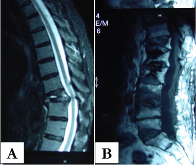
Figure 5.

A and B, Magnetic resonance imaging (MRI) of a patient with vertebral compression fracture. Note the marrow edema and increased signal on the T2 image. One should also evaluate the MRI for other areas of involvement. (Images courtesy of Dr Howard Place.)
