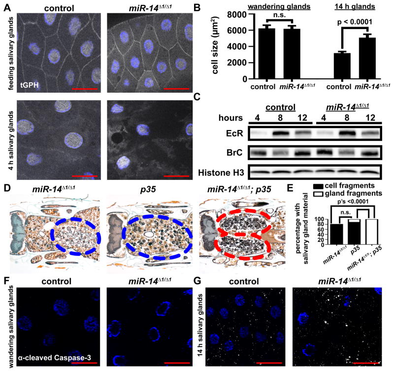
Figure 2. Loss of miR-14 does not alter cell growth, hormone signaling, or caspase activity during salivary gland degradation.
(A) Salivary glands dissected from control (+/w; +/miR-14Δ1, tGPH) and miR-14 null mutant (w; miR-14Δ1/miR-14Δ1, tGPH) animals as feeding larvae (top) and 4 h after puparium formation (bottom) for analysis of tGPH localization. Salivary glands were all stained with Hoechst (blue). Scale bars represent 50μm.
(B) Salivary gland cell size in control (+/w; +/miR-14Δ1), n ≥ 32, and miR-14 null mutant (w; miR-14Δ1/miR-14Δ1), n ≥ 29, animals dissected from wandering larvae and 14 h after puparium formation. Statistical significance was determined using a Student’s t-test. Data are represented as mean +/− SEM.
(C) Protein extracts from salivary glands 4, 8 and 12 hours after puparium formation of control (+/w; +/miR-14Δ1) and miR-14 null mutant (w; miR-14Δ1/miR-14Δ1) animals analyzed by immuno-blot with anti-Ecdysone Receptor (EcR) and anti-Broad Complex (BR-C).
(D) Samples from miR-14 null mutants (w; miR-14Δ1/miR-14Δ1; UAS-p35/+), n = 20 (left), animals with salivary gland-specific expression of p35 (w; miR-14Δ1/+; UAS-p35/fkh-GAL4), n = 26 (middle), and miR-14 null mutants with salivary gland-specific expression of p35 (w; miR-14Δ1/miR-14Δ1; UAS-p35/fkh-GAL4), n = 24 (right), analyzed by histology for the presence of salivary gland material 24 h after puparium formation. Blue dotted circles enclose cell fragments; red dotted circles enclose gland fragments.
(E) Quantification of data from (D). Data are represented as mean. Statistical significance was determined using a Chi-squared test comparing the percentages of gland fragments. (F and G) Salivary glands dissected from control (+/w; +/miR-14Δ1) and miR-14 null mutant (w; miR-14Δ1/miR-14Δ1) animals and stained with cleaved Caspase-3 antibody (white) and Hoechst (blue). Salivary glands were dissected from wandering larvae (F) and 14 h after puparium formation (G). Scale bars represent 50 μm.

