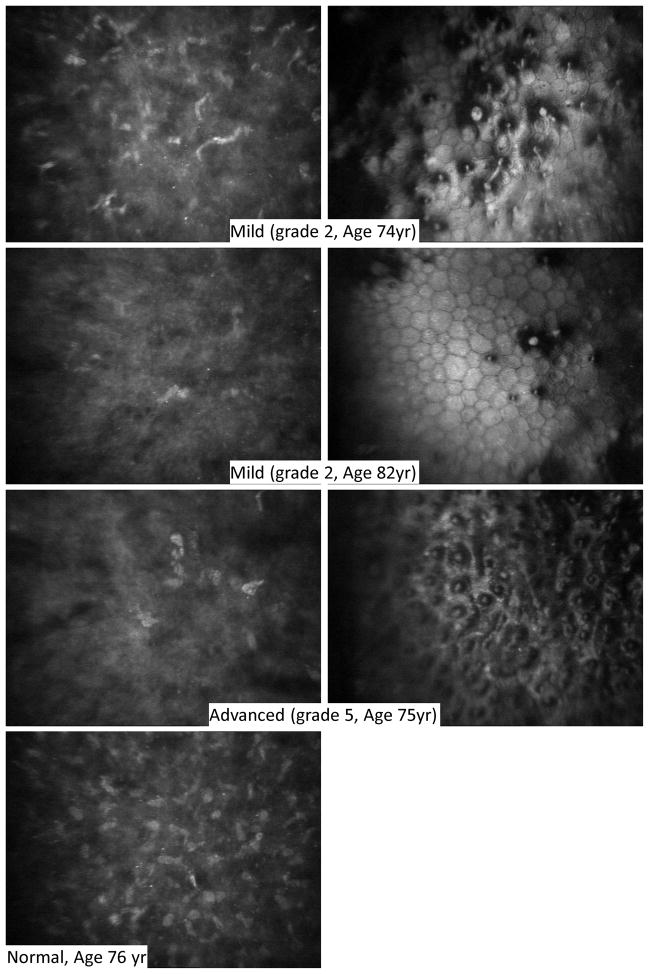
Figure 4.
Anterior stromal cells and associated endothelium in Fuchs dystrophy. The most anterior stromal cells (left) were depleted and extracellular reflectivity was higher in Fuchs dystrophy (upper three rows) compared to normal (bottom row). Anterior stromal cell depletion was evident even in mild cases of Fuchs dystrophy (upper two rows) in which the corresponding endothelial images (right) have scattered guttae. One case of advanced Fuchs dystrophy (third row) is also shown with depleted anterior stromal cells (left) and confluent guttae (right).

