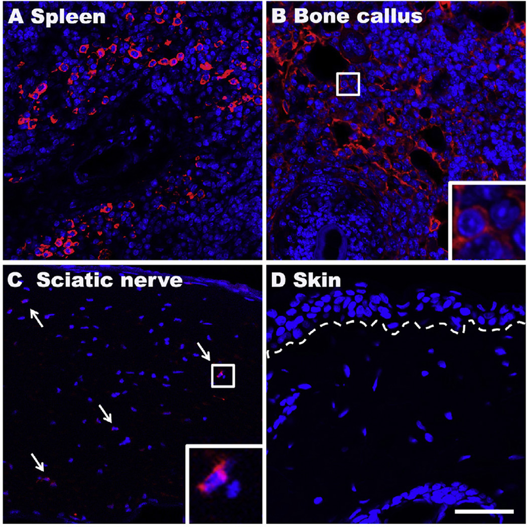
Fig. 7.
Representative fluorescent photomicrographs of immunostaining for the B-cell marker CD20 (red) at 3 weeks postfracture. DAPI (blue) counterstain was performed to show DNA content and nuclei. Abundant CD20 (cytoplasmic/red) positive B cells were observed in spleen (A) and bone callus near the site of fracture (B), and scattered B cells in sciatic nerves (C), but not seen in skin (D). Dotted line indicates the epidermal-dermal boundary, arrows: B cells, small boxed regions in panels (B) and (C) are shown enlarged in the lower right corner of the panels, respectively; scale bar = 50 µm.

