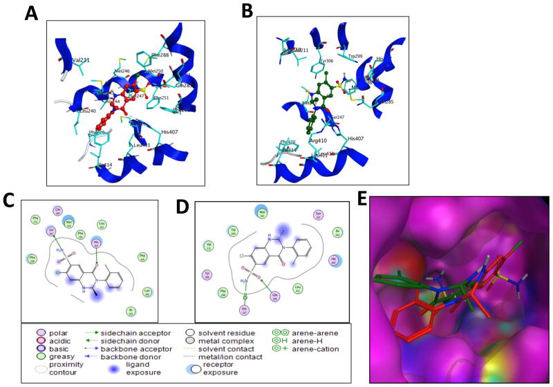
Fig. 9. Binding orientations of MET at the PXR LBD binding site.
(A) Docking mode three-dimensional (3D) interaction scheme of predicted binding orientations of MET (shown in red) at the PXR LBD binding site (Orientation 1). (B) Docking mode 3D interaction scheme of another predicted binding orientation of MET (shown in green) at the PXR LBD binding site (Orientation 2). (C) Docking mode two-dimensional (2D) interaction scheme of predicted binding pose of MET at the PXR LBD binding site (Orientation 1). (D) Docking mode 2D interaction schemes of predicted binding pose of MET at the PXR LBD binding site (Orientation 2). (E) Surface representation of the PXR LBD bound to MET in a different orientation. MET orientations are shown in stick with green and red color.

