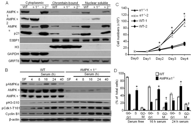
Fig. 1.
AMPKα1 localizes to chromatin and is implicated in regulating the cell cycle. (A) MEFs were subcellularly fractionated by a commercially available kit from Thermo Scientific. AMPKα1, AMPKα2, AMPKα, p21, and 53BP1 were detected by Western blots. Histone H3 serves as a marker for chromatin. GAPDH serves as a cytoplasmic marker. GRP78 serves as a loading control for each fraction. Representative data from three independent experiments are shown. (B) AMPKα is activated during mitosis. WT and AMPKα1−/− MEFs were first serum-deprived for 24 hours, then cultured in regular culture medium for the indicated times. The cells were lysed and analyzed by Western blot using anti-pAMPKα-T172, -AMPKα, -AMPKα1, -Cyclin B1, -pCdk1-T161, or pH3-S10 antibody. Representative data from three independent experiments are shown. (C) WT and AMPKα1−/−-immortalized MEFs (two independent cell lines for each) were plated, and cells were counted at the indicated times. n=8, *p < 0.05 vs WT. (D) Flow cytometric analysis of cell cycle progression in WT and AMPKα1−/− MEFs serum starved for 24 hours or treated with serum for the indicated times. n=3, *p < 0.01 vs WT in S phase; † p < 0.01 vs WT in G2-M phase.

