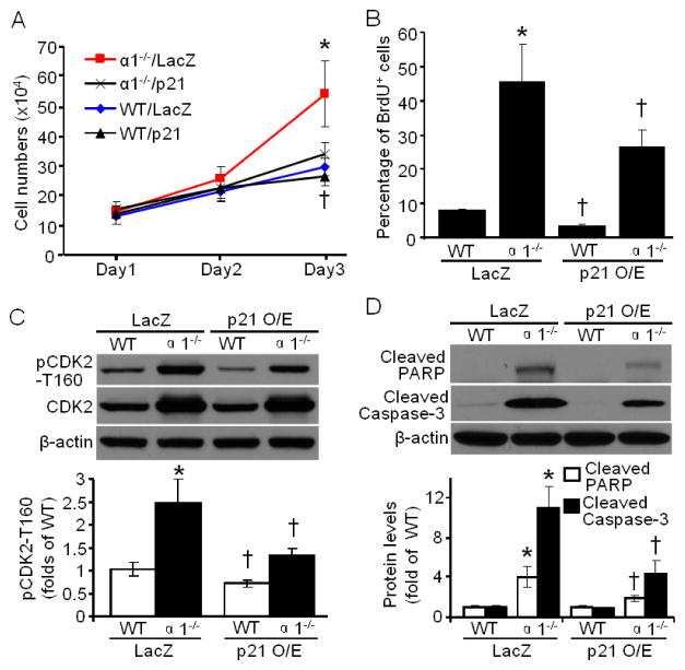
Fig. 6.
p21 overexpression attenuates cellular hyperproliferation and apoptosis in AMPKα1−/− MEFs. (A) p21 overexpression blocked the hyperproliferation of AMPKα1−/− MEFs. n=10, *p < 0.01 vs α1−/−/p21; † p < 0.05 vs WT/LacZ. (B) p21 overexpression decreased the percentage of BrdU positive cells in AMPKα1−/− and WT MEFs. n=4, *p < 0.01 vs WT/LacZ; †p < 0.01 vs WT/LacZ, α1−/−/LacZ, respectively. (C) (Upper) p21 overexpression significantly inhibited phosphorylation of CDK2 at T160 (pCDK2-T160), while had no effect on total CDK2 protein level. (Bottom) Quantification of Western blot data. n=3, *p < 0.001 vs WT/LacZ; † p < 0.01 vs WT/LacZ, or α1−/−/LacZ, respectively. (D) p21 overexpression suppressed apoptotic signal (PARP and Caspase-3 cleavage) in AMPKα1−/− MEFs. n= 3, *p < 0.001 vs WT/LacZ; † p < 0.01 vs α1−/−/LacZ.

