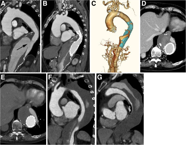
Figure 2.

A 78-year-old female with type B chronic AD. A. Six years after onset, contrast-enhanced CT image shows aneurysmal dilation of the affected aorta and entry site (arrow). B, C. TEVAR (GORE TAG: 37-mm diameter & 20-cm length) was performed. Contrast-enhanced CT image and 3D CT angiography show that the entry site was closed. D, E. Six (F) and (G) 15 months after TEVAR, contrast-enhanced CT images show that the false lumen has partially thrombosed, but aneurysmal dilation of the affected aorta remains.
