
Figure 1.
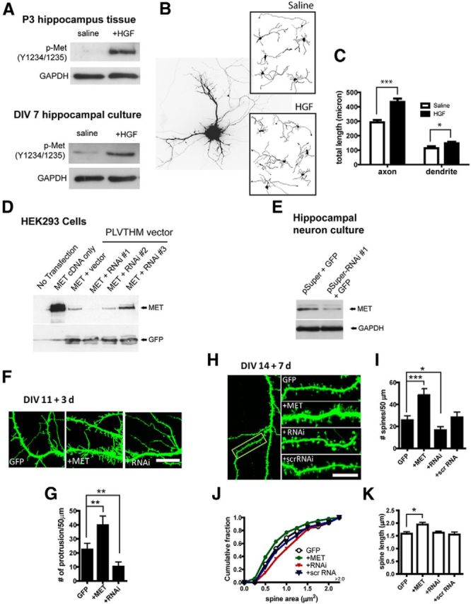
MET signaling affects growth and morphological development of hippocampal neurons in vitro. A, MET signaling competency in live P3 hippocampus slices and DIV 7 cultured hippocampal neuron. HGF stimulation leads to tyrosine (Y1234/1235) phosphorylation of MET. B, Cultured hippocampal neurons were transfected with GFP to reveal morphology. HGF treatment enhances the growth of both developing dendrites and axons. C, Quantification of neurite growth as shown in B (* p < 0.05; ***p < 0.001). D, Test of efficacy of three 19-nt RNAi sequences by cotransfection with MET cDNA in HEK293 cells. RNAi sequences were cloned into PLVTHM vector, which has bicistronic expression of GFP. RNAi #1 sequence is most efficient in reducing MET expression. E, Test of knockdown efficiency of RNAi sequence #1 on endogenous MET expression in cultured hippocampal neurons. This RNAi sequence was cloned into pSuper vector for faster expression and designated as ‘RNAi’ hereafter. F, MET OE and RNAi affect dendritic protrusions of cultured hippocampal neurons during the second week in culture. G, Quantification of F. MET OE significantly increased (**p < 0.01), while RNAi significantly decreased (**p < 0.01), the density of dendritic protrusions. H–K, MET OE or RNAi alters dendritic spine density and morphology (H). MET OE increases (***p < 0.001), while RNAi decreases (*p < 0.05), the density of dendritic spines (I). A scrambled sequence of RNAi was without effect. MET OE decreases, while RNAi increases, spine head area (J) (p < 0.01, for both effects). MET OE also significantly increases dendritic spine length (K) (*p < 0.05). Scale bars: F, H, 10 μm.