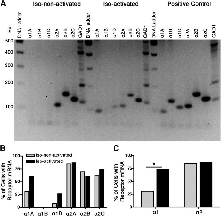
Figure 3.
Qualitative multiplex RT-PCR showed a difference in α1 adrenergic receptor expression between isoflurane-activated and isoflurane-non-activated VLPO neurons. A, Representative gel revealing the expression of α-adrenergic receptors and GAD1 PCR products from an isoflurane-non-activated neuron, an isoflurane-activated neuron, and whole-brain library cDNA positive controls. B, Percentage of α-adrenergic receptor subtypes detected in isoflurane-non-activated neurons (gray) and isoflurane-activated neurons (black). C, Percentage of each cell type containing any subtype of α1 receptors and α2 receptors. Data were analyzed by χ2 test, *p < 0.05.

