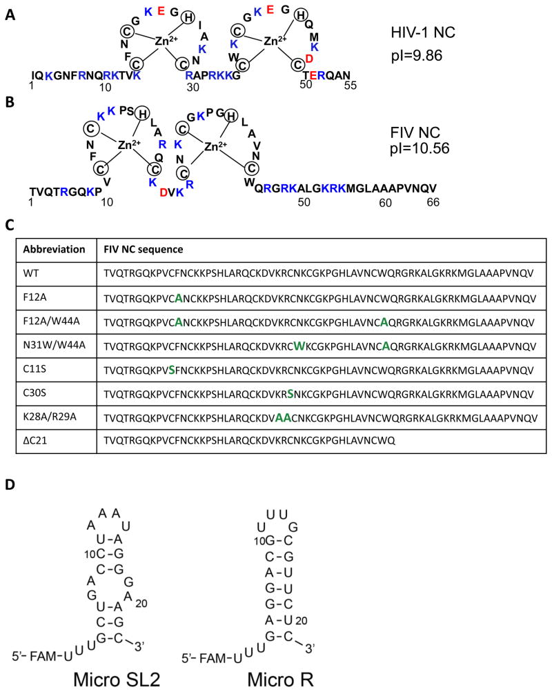
Fig. 1.
(A) Sequence of WT HIV-1 NC (NL4-3 isolate). (B) Sequence of WT FIV NC (MD isolate). Positively and negatively charged residues are highlighted in blue and red, respectively, in both (A) and (B). (C) Sequence of WT and mutant FIV NC, with altered residues highlighted in green. (D) Predicted secondary structures of micro SL2 and micro R RNAs derived from the FIV genome. Three U’s that are not encoded in the genome and a 5′-FAM label are also indicated.

