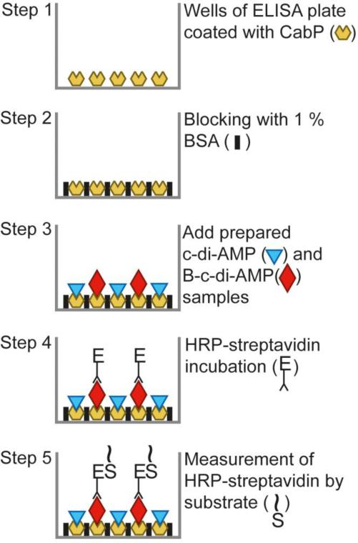
Fig. 1.
Diagrammatic representation of the competitive ELISA for detection of c-di-AMP. Symbols are defined where appropriate. The first part of the assay relies on the coating of wells with CabP protein followed by a standard blocking step with BSA. A mixture of biotin-labeled and unlabeled c-di-AMP in standards or samples is then added to create the competitive condition in each well. Finally, the bound B-c-di-AMP is detected by incubation with HRP-conjugated streptavidin and followed by reaction with an OPD substrate.

