Figure 1.
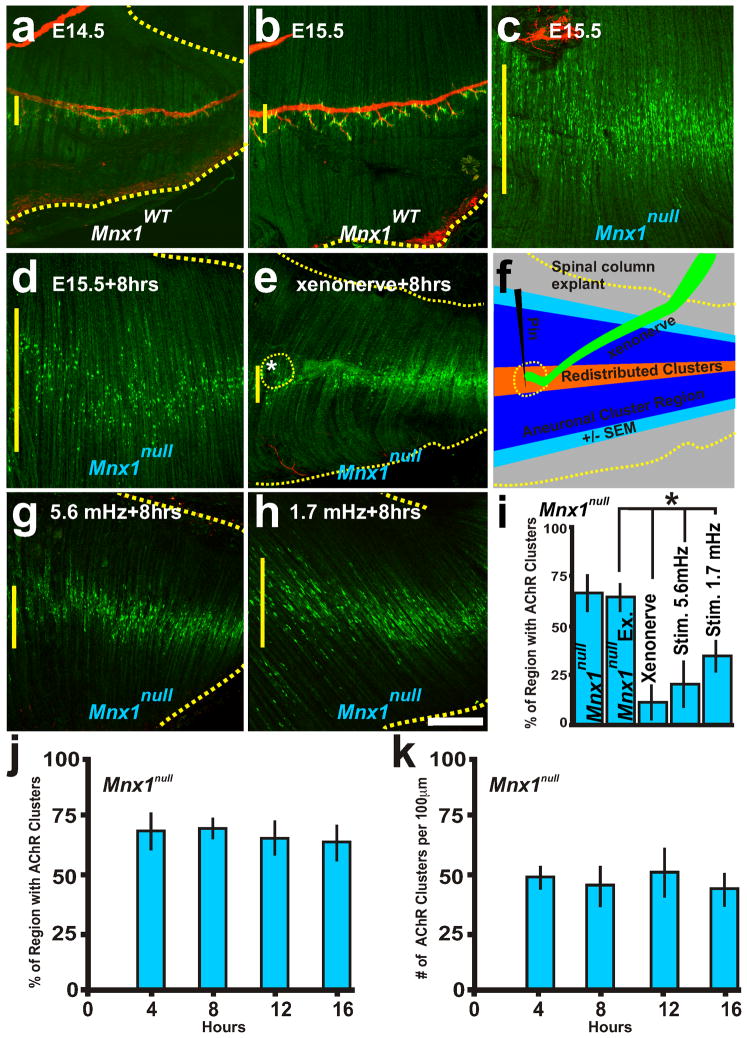
Neuronal and electrical activity cues control the distribution of AChR clusters. Representative examples of AChR clusters (α-bungarotoxin conjugated Alexa-488; green) and the phrenic nerve (neurofilament-160; red). a, b, E14.5 and E15.5 diaphragm from wildtype mice with AChR clusters apposed to the phrenic nerve. c, E15.5 diaphragms from Mnx1null mice without a phrenic nerve. d, E15.5 Mnx1null diaphragm explant cultured for 8 hours. e, Littermate Mnx1null diaphragm with Mnx1GFP/+ spinal cord-phrenic nerve explant pinned during 8 hr culture (asterisk and dotted yellow line indicate xenonerve placement, the xenonerve detached during fixation) shows a narrowed AChR cluster region compared to d. f, Schematic of explant showing the region of aneural AChR clusters at the start of the experiment (blue) and the redistribution and centralization of AChR clusters in the presence of the xenonerve (orange). g, h, E15.5 Mnx1null diaphragms stimulated every 3 minutes (5.6 mHz) or 10 minutes (1.7 mHz) for 8 hours induces redistribution and elimination of AChR clusters. i, Quantification of the distribution of AChR clusters within the Mnx1null explant diaphragms before and after culture, with xenonerve, and following electrical stimulation. j, k, Percent regionalization (j) and number (k) of AChR clusters at 4, 8, 12, and 16 hours of culture of Mnx1null diaphragm explants. Scale bar = 200 μm; yellow vertical line on the left of each panel represents the largest width of the AChR cluster region within ventral quadrant of diaphragm shown. Each condition was examined in 5 diaphragms.

