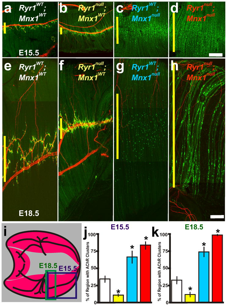
Figure 4.
Nerve and muscle components are required for centralization of AChR clusters. a–d, E15.5 and e–h, E18.5 composite pictures of diaphragm muscle from RyR1WT; Mnx1WT (white lettering and box in j,k), RyR1null; Mnx1WT (yellow), RyR1WT; Mnx1null (blue) and RyR1null; Mnx1null (red). i, Illustration of diaphragm muscle and regions analyzed at E15.5 (purple box) and E18.5 (green rectangle representation of composite picture in f). Purple rectangle represents E15.5 images and regions of quantification presented in figure graphs at E15.5. j, E15.5 graph of the percent of diaphragm with patterned AChR clusters in all diaphragm crosses examined in ad. k, E18.5 graph of the percent of diaphragm with patterned AChR clusters in all diaphragm crosses examined in e–h excluding random AChR clusters in RyR1null; Mnx1WT diaphragm muscles (n=5 per cross). Asterisks show significance (p<0.05) of samples compared to control. Scale bar for a–d=200μm; for e–h= 50 μm. a, c are from Figure 1b,c. Yellow vertical line on the left of each panel represents the largest width of the AChR cluster region within ventral quadrant of diaphragm shown. Each condition was performed in 6 diaphragms.

