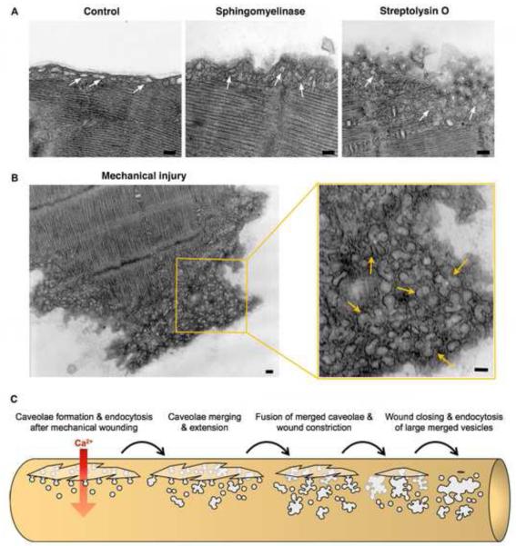
Figure 3. Caveolae-like vesicles accumulate below the sarcolemma of primary muscle fibers in response to injury or exposure to purified sphingomyelinase.
(A) Transmission EM images of mouse Flexor digitorum brevis muscle fibers untreated (Control), exposed to 50 mU/ml B. cereus sphingomyelinase for 5 min, or treated with 400 ng/ml SLO for 30 seconds. (B) Transmission EM image of the mechanically severed tip of a Flexor digitorum brevis muscle fiber. Arrows in the enlarged section to the right point to merged caveolae-derived compartments below the sarcolemma. Bars= 100 nm. (C) Proposed caveolae-mediated mechanism for the resealing of mechanical wounds on a muscle fiber. Ca2+ flowing through a wound would induce internalization and intracellular merging of sarcolemma-associated caveolae, leading to the formation of larger compartments tethered to the sarcolemma that constrict and ultimately reseal the wound.

