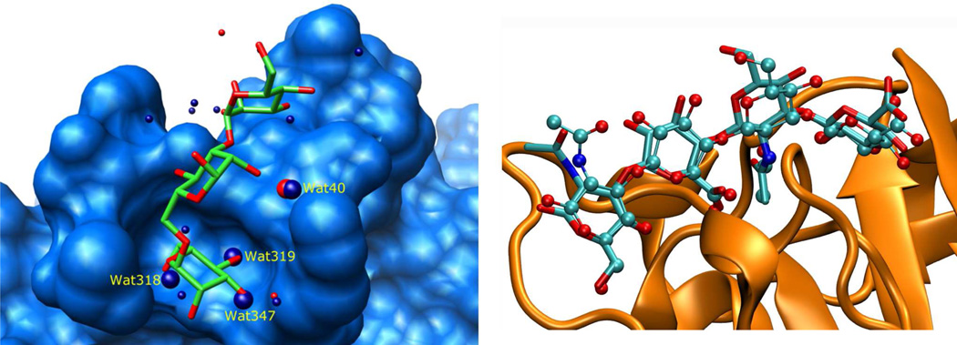
Figure 3.
Left: solvent accessible surface of the lentil lectin Concanavalin A (light blue) in complex with the natural trisaccharide ligand (PDB: 1CVN). Waters of crystallization are shown as red spheres. Superimposed are the waters of crystallization (dark blue) from the ligand-free structure (PDB: 1GKB). Key waters, which appear to be displaced by the ligand, as well as a conserved water molecule (Wat40), are shown as larger spheres [57]. Right: results from docking a hyaluronan tetrasaccharide to CD44 (orange, ribbon model) using the water-site biased docking method. The top ranked pose (ball and stick model) has an RMSD of 0.7 Å from the reference ligand position (stick model) [63].

