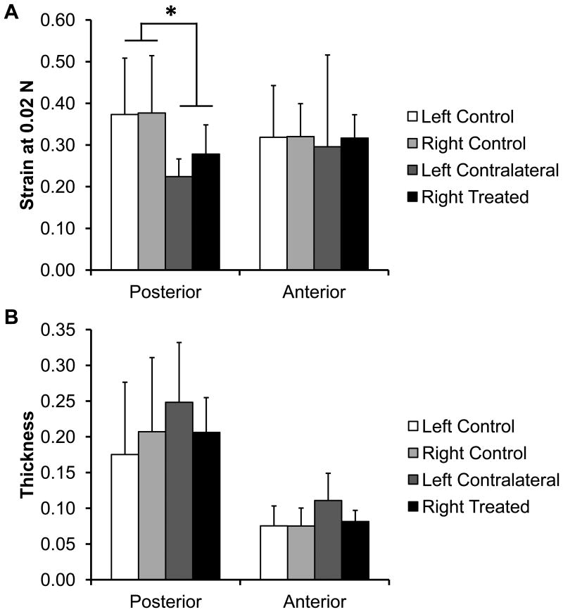
Figure 6.
Condylar fibrocartilage compression results. A) Strain of the condylar fibrocartilage at 0.02 N of loading, as compression testing to a particular force was used on the TMJ condyles regionally. The posterior region of the splinted rabbit condyles was significantly stiffer than the control fibrocartilage. (*p>0.05) No other statistically significant differences were observed. B) Thickness of the TMJ condylar fibrocartilage. No statistically significant differences were observed. Sample size of n=8 per group. All error bars are standard deviations.

