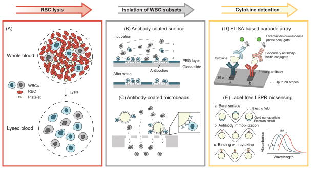
Figure 2.
Paradigm of functional immunophenotyping of WBCs using microengineering tools. Background RBCs are first removed from whole blood samples by lysis. Target WBC subsets are then isolated for cytokine secretion measurements. (A) Schematic of RBC lysis. (B) Antibody-coated surface for capturing subsets of WBCs. Target WBCs are bound to the antibodies coated on the glass slide during incubation, while unbounded cells are washed away [36]. (C) Isolation of subsets of WBCs using antibody-coated microbeads and microfilters. Target WBCs are retained on the filter as the microbead is larger than the filter pore size, while all other undesired WBCs pass through the filter freely [48]. (D) High-density antibody barcode using sandwich ELISA for multiplexed detection of secreted proteins [44, 45]. Adapted with permission from [44]. (E) Principle of LSPR biosensing on gold nanoparticle surface. The absorbance spectrum peak shift is used to quantify the amount of cytokine secreted by WBCs [51]. Adapted with permission from [51].

