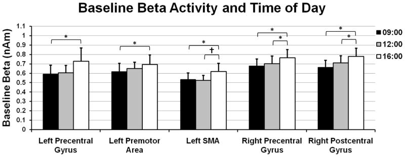
Figure 6.

Beta activity levels during the baseline period (i.e., before movement) also increased from 09:00 to 16:00 in sensorimotor cortices. To evaluate time-related changes in baseline beta activity, we extracted virtual sensors from the peak voxels of the beta ERD response and computed the mean beta amplitude during the baseline period. The results indicated a significant linear increase in the amplitude of baseline beta activity across time (p < 0.05) in the left precentral gyrus, left SMA, left premotor cortices, and the right precentral and postcentral gyri. Error bars indicate one standard error of the mean. Significant differences based on pairwise comparisons have been marked (* = p < 0.05; † = p < 0.10).
