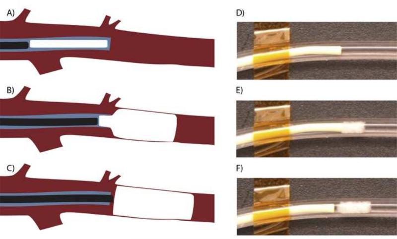
Figure 4.
Schematic diagram of endovascular deployment of the SMP foam VOD: (A) the device is pushed near the 5F catheter tip by the guidewire, (B) the guidewire pushes the self- actuating device out of the catheter, and (C) the deployed device fills the vessel lumen. (D-F) In vitro demonstration of VOD deployment showing immediate expansion of the VOD in 37°C (body temperature) water in a silicone tube (3.5 mm inner diameter).

