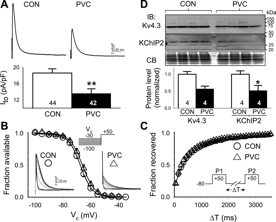
Fig. 2. PVC myocytes had reduced transient outward (Ito) current density but unaltered Ito gating kinetics, accompanied by a downregulation of Ito channel subunits.
(A) Top: Representative Ito traces recorded from CON and PVC myocytes. Bottom: peak Ito densities in CON and PVC myocytes. (B) Voltage-dependence of Ito inactivation. (C) Time course of recovery from inactivation (Ito restitution). (D) Immunoblot quantification of protein levels of pore-forming and auxiliary subunits of Ito channels (Kv4.3 and KChIP2). Top: Images of immunoblots and Coomassie blue stain of the gel (CB, as loading control). Size marker bands (in kDa) are shown on the right. Bottom: Densitometry quantification of Kv4.3 and KChIP2 protein levels.

