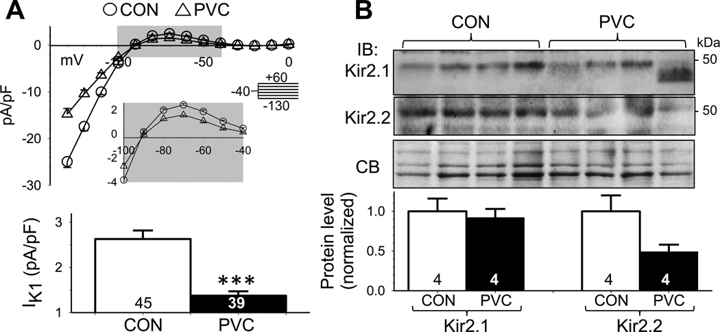
Fig. 3. PVC myocytes had reduced inward rectifier (IK1) current densities, accompanied by a downregulation of Kir2.2.
(A) Top: Average steady-state current-voltage (I-V) relationships of CON and PVC myocytes. The enlarged view of gray shading area highlights the outward portion of IK1. Bottom: outward IK1 densities at −70 mV in CON and PVC myocytes. (B) Immunoblot quantification of IK1 channel subunits, Kir2.1 and Kir2.2, in LV samples. Data analysis and presentation are the same as Fig. 2D.

