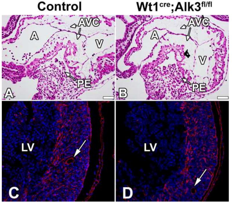
Figure 3. Proepicardium formation and EPDC contribution to the LV is unaffected in Wt1cre;Alk3fl/fl;R26mG hearts.

H/E stained sections of ED9.5 control (A) and Wt1cre;Alk3fl/fl (B) specimens show normal formation of the proepicardium. ED17 control (C) and Wt1cre;Alk3fl/fl;R26mG (D) specimens immunolabeled for DAPI (blue) and eGFP (red) demonstrate that EPDC contribution to the LV is unaltered in the Wt1cre;Alk3fl/fl;R26mG hearts. Arrows indicate EPDC contribution to the coronary vasculature. (Scale bar = 50μm). A, atria; AVC, atrioventricular cushion; LV, left ventricle; PE, proepicardium; V, ventricle.
