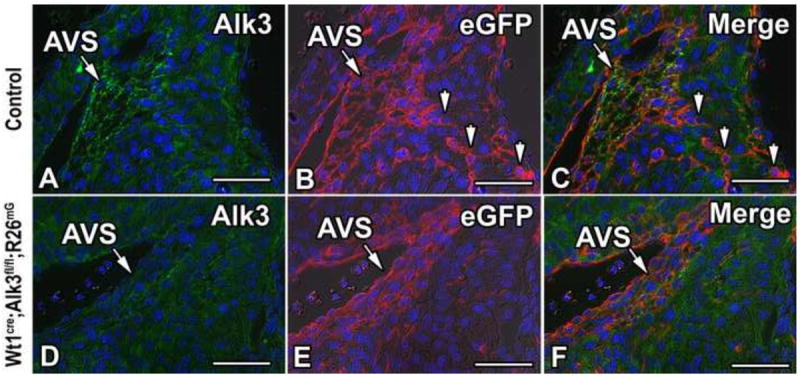
Figure 4. Loss of Alk3 expression in the AV sulcus of Wt1cre;Alk3fl/fl;R26mG mice.

Immunolabeling for Alk3 (green, A, C, D, F), eGFP (red, B, C, E, F), and DAPI (blue, A- F) in the right AV junction of ED13 control (A-C) and Wt1cre;Alk3fl/fl;R26mG (D-F) specimens. In panel C, the channels depicted in A and B are merged, while in panel F the channels depicted in D and E are merged. Panels A-C show that Alk3 is expressed in the eGFP labeled EPDCs of the AV sulcus. Panels D-F show that in Wt1cre;Alk3fl/fl;R26mG mice Alk3 is efficiently deleted from the EPDCs in the AV sulcus. Arrowheads in B, C indicate eGFP labeled EPDCs within the AV canal myocardium of the control specimen. (Scale bar = 50μm). AVS, AV sulcus.
