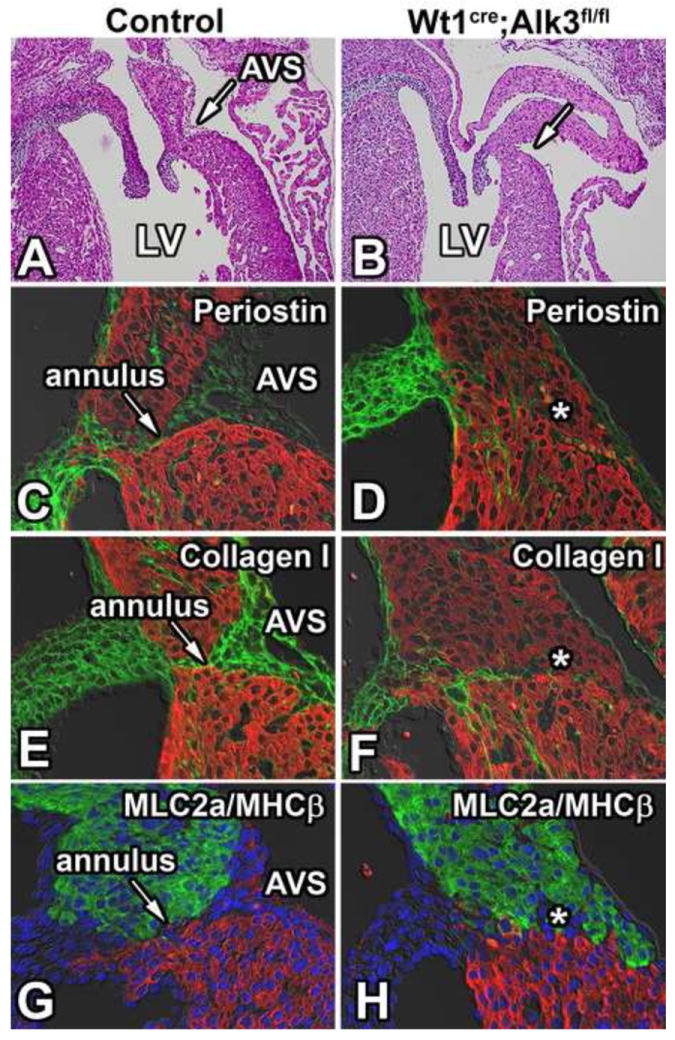
Figure 6. Loss of Alk3 in EPDCs results in malformation of the annulus fibrosus.

Panels A and B show the left AV junction of H/E stained sections of ED17 control and Wt1cre;Alk3fl/fl;R26mG hearts. The AV sulcus is absent in the left AV junction of the Wt1cre;Alk3fl/fl;R26mG heart (B). Immunolabeling for MF20 (red, C-F), periostin (green; C,D) and collagen I (green; E,F) in these specimens shows that the annulus fibrosus, present in the control heart (arrow in C,E,G), is absent in the Wt1cre;Alk3fl/fl;R26mG heart (asterisk in panels D, E, and H). Immunolabeling for MLC2a (green; G,H), MHCβ (red; G,H), and DAPI (blue; G,H) marks where the atrial (MLC2a) and ventricular myocardium (MHCβ) meet in the AV junction. AVS, AV sulcus; LA, left atria; LV, left ventricle.
